
No products in the cart.
Articles
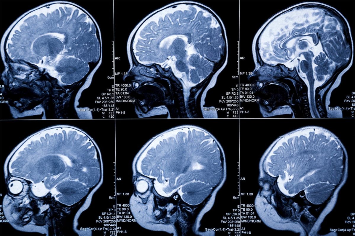
New MRI Technique Might Help Spot MS Sooner
07
Jan
Jan
By Cara Murez
HealthDay Reporter
FRIDAY, Jan. 7, 2022 (HealthDay News) – Researchers in Austria say a brand new MRI method might result in quicker prognosis and remedy for folks with a number of sclerosis.
The method can detect biochemical adjustments within the brains of individuals with MS early of their illness, in accordance with findings printed Jan. 4 within the journal Radiology.
“MRI of neurochemicals enables the detection of changes in the brain of multiple sclerosis patients in regions that appear inconspicuous on conventional MRI,” stated examine senior creator Wolfgang Bogner, from the High Field MR Centre on the Medical University of Vienna. “The visualized changes in neurochemistry of normal-appearing brain tissue correlated with the patients’ disabilities.”
MS, a disease of the central nervous system, affects nearly 3 million people worldwide. There is no cure, and it can cause fatigue, pain and impaired coordination. Physical therapy and medications can slow its progression.
Currently, MS can be detected in lesions in the brain’s white matter on standard MRIs. These lesions are linked to the loss of a protective coating around nerve fibers known as myelin. This is tissue damage visible to the naked eye but finding the damage when it is still microscropic or at a biochemical stage would be better.
An advanced imaging technique, called proton MR spectroscopy, can detect substances produced during metabolism that have potential relevance for MS, the researchers say.
They used this to compare biochemical changes in the brains of 65 people with MS with those of 20 healthy individuals. They employed an MRI scanner with a powerful 7-Tesla (T) magnet.
The team found reduced levels of an amino acid derivative called N-acetylaspartate (NAA) in patients with MS. Lower levels of NAA have been linked to impaired integrity of neurons in the brain.
People with MS also showed elevated levels of myo-inositol (MI), a compound involved in cell signaling. Higher levels can indicate substantial inflammatory disease activity.
Researchers said the results show a potential role for the new MRI technique in visualizing MS pathology beyond demyelinating lesions.
“Some neurochemical changes, particularly those associated with neuroinflammation, occur early in the course of the disease and may not only be correlated with disability, but also be predictive of further progression such as the formation of multiple sclerosis lesions,” stated examine lead creator Eva Heckova, additionally from the High Field MR Centre. She stated the adjustments detected by this new imaging method might have vital medical functions.
More work is required to substantiate the outcomes, nevertheless.
“If confirmed in longitudinal clinical studies, this new neuroimaging technique could become a standard imaging tool for initial diagnosis, for disease progression and therapy monitoring of multiple sclerosis patients and, in concert with established MRI, might contribute to neurologists’ treatment strategies,” Bogner stated in a journal information launch.
More info
The U.S. National Library of Medicine has extra on a number of sclerosis.
SOURCE: Radiological Society of North America, information launch, Jan. 4, 2022